
Applications of fluorescence-guided surgery in head and neck surgery – a narrative review
Authors:
M. Kaňa
 ; J. Plzák
; M. Zábrodský
; J. Bouček
; J. Plzák
; M. Zábrodský
; J. Bouček
Authors place of work:
Homolka, Prague
; Department of Otorhinolaryngology and Head and Neck Surgery, 1st Medical Faculty, Charles University and University Hospital Motol and
Published in the journal:
Otorinolaryngol Foniatr, 75, 2026, No. 3, pp. 259-270.
Category:
Přehledový článek
doi:
https://doi.org/10.48095/ccorl2026-011
Summary
Fluorescence-guided surgery (FGS) has emerged as a valuable intraoperative imaging technique that enhances the real-time visualization of anatomical structures and pathological tissues. In head and neck surgery, where the anatomical complexity and proximity of vital structures pose significant challenges, FGS enhances surgical precision and outcomes. This review provides a comprehensive overview of clinical applications, technical aspects, and recent advances in fluorescence-guided surgery within the field of otolaryngology and head and neck surgery. FGS has demonstrated significant utility in identifying critical anatomical structures such as nerves, vasculature, and lymphatics; and evaluating flap perfusion. Its role in thyroid and parathyroid surgery is well established, where it aids in preserving gland function and reducing postoperative complications. Recently, its applications in oncosurgery, particularly insitu tumor margins assessment, ex vivo evaluation of resected specimens, and sentinel lymph nodes mapping, are rapidly evolving. Even though the number of clinically approved fluorophores remains limited, the growing number of ongoing clinical trials indicates a strong potential for further expansion of this technology. FGS represents a valuable tool in modern head and neck surgery, improving intraoperative visualization, enhancing surgical safety, and supporting oncologic and functional outcomes.
Keywords:
lymph node – head and neck cancer – parathyroid gland – surgical margins – fluorescence-guided surgery – near-infrared imaging
Introduction
Fluorescence-guided surgery (FGS) is an intraoperative imaging technique that enables real-time visualization of anatomical structures and differentiation of pathological tissues. In recent years, it has gained considerable attention in head and neck surgery, where surgical precision is critical due to the dense anatomical complexity and the proximity of vital structures. The clinical rationale for FGS lies in its ability to enhance intraoperative contrast between normal and diseased tissues, support functional preservation, and potentially improve oncological outcomes by assisting in complete tumor resection [1, 2].
The principle of FGS relies on the use of fluorophores – molecules that emit fluorescence upon excitation by the light of a specific wavelength. Once excited, these molecules return to their ground state by releasing photons, producing a fluorescent signal detectable with specialized imaging systems.
Fluorescence can originate either from intrinsic tissue components – known as autofluorescence – or from exogenous probes administered to the patient prior to surgery. Autofluorescence, arising from molecules such as NADH, collagen, or elastin, can help differentiate tissue types, but it often suffers from low specificity and limited contrast in the surgical setting [3]. Therefore, the majority of clinical FGS applications rely on exogenous fluorescent probes, which are engineered for enhanced signal-to--background ratios and greater tissue selectivity. These can be either non-specific agents, such as indocyanine green (ICG), or targeted agents conjugated to antibodies, peptides, or small molecules specific to tumor biomarkers [1–3].
The most widely used and clinically approved fluorophores include ICG, fluorescein sodium, 5-aminolevulinic acid (5-ALA) and methylene blue. ICG currently as the most widely used fluorescent probe accumulates in tissues based on perfusion and is commonly used for angiography and sentinel lymph node mapping. Its emission in the NIR-I window (near-infrared, 700–900 nm) allows for improved tissue penetration and reduced background noise from autofluorescence, which is more prominent at shorter (visible) wavelengths [4]. Fluorescein, emitting in the visible spectrum, has been applied in ophthalmic and neurosurgical procedures, while 5-ALA, metabolized to protoporphyrin IX, is used for glioma visualization [5, 6].
In the field of head and neck surgery, particular emphasis has been placed on targeted fluorescent agents, especially monoclonal antibodies such as cetuximab and panitumumab conjugated with IRDye800CW. These agents target the epidermal growth factor receptor (EGFR), which is overexpressed in a large proportion of head and neck squamous cell carcinomas (HNSCC). Clinical trials have demonstrated that EGFR-targeted tracers such as cetuximab-IRDye800CW significantly improve intraoperative identification of tumor margins, lymph node metastases, and residual disease, with reported sensitivities and specificities exceeding 90% [7–9]. This high degree of accuracy facilitates more complete resections and minimizes damage to surrounding healthy tissue.
Beyond antibodies, other classes of probes include small peptides, activatable probes, and hybrid agents designed for multimodal imaging. Activatable probes remain optically silent until cleaved or modified by specific enzymes within the tumor microenvironment, increasing specificity while reducing background signal [4, 5]. Hybrid imaging agents combine fluorescence with nuclear medicine or magnetic resonance modalities, enabling complementary preoperative imaging (e. g., PET or MRI) and real-time intraoperative fluorescence visualization [2, 7, 9, 10].
The success of FGS also hinges on the availability of sophisticated clinical imaging systems capable of detecting fluorescence signals against the complex background and visualize fluorescence in real time. In current clinical practice, several systems have been adopted and validated across surgical disciplines, including open-field, robotic-integrated, and closed-field platforms. These systems combine high-sensitivity cameras (typically CMOS or CCD), optimized excitation light sources, and wave--length-specific emission filters to enable real-time overlays of fluorescence on standard white-light images [1, 8]. Among the most widely used platforms is the SPY-PHI® system (Stryker), commonly employed for open surgical visualization of ICG fluorescence in perfusion and lymphatic mapping [11]. The VisionSense VS3® system (Medtronic) integrates three-dimensional visualization with NIR fluorescence and has been applied in skull base and head and neck oncologic procedures [12, 13]. In the context of robotic procedures, the Firefly® imaging system, integrated into the da Vinci® Surgical System (Intuitive Surgical), allows intraoperative NIR fluorescence imaging during transoral robotic surgery (TORS) and neck dissections [14, 15]. This FDA-approved system supports ICG imaging and has been widely adopted due to its seamless integration and intuitive interface.
Additionally, closed-field systems such as the PEARL Trilogy® (LI-COR) and Fluobeam® (Fluoptics) are used for ex vivo imaging of resected specimens and for parathyroid gland autofluorescence assessment [16, 17].
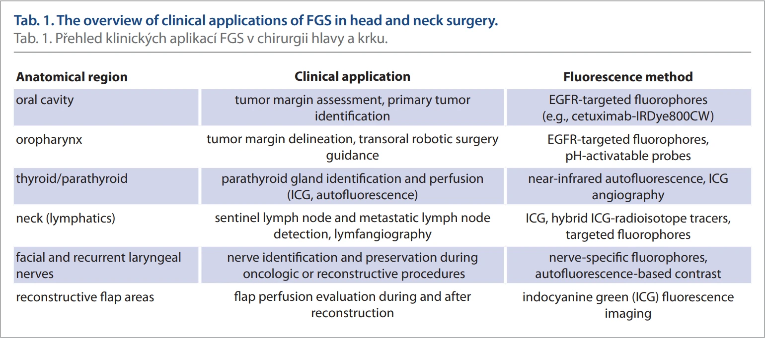
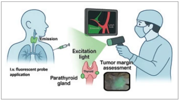
In head and neck surgery, where precision is critical due to the dense anatomical complexity and proximity of vital structures, the FGS offers high transformation potential (Tab. 1). As the technology continues to evolve, the integration of FGS with other intraoperative tools – such as Raman spectroscopy, photoacoustic imaging, and augmented reality – is expected to further enhance surgical outcomes by improving tissue discrimination and reducing subjectivity in visual assessments (Fig. 1) [18, 19].
Methods
A literature search was conducted on PubMed, Scopus and Web of Science databases using specific keywords: (“fluorescence-guided” OR “fluorescence-navigated” OR “fluorescence imaging” OR “near-infrared fluorescence” OR “NIR fluo-rescence” OR “fluorescence navigation”) AND (“head and neck” OR “otolaryngology”) AND (“head and neck surgery” OR “head and neck cancer” OR “oral cancer” OR “oropharyngeal” OR “thyroid” OR “parathyroid” OR “larynx” OR “salivary gland” OR „squamous cell carcinoma” OR “nerve imaging” OR “angiography“ OR “lymphatic”). The search was performed on 26th March 2025, no limitation on publication date was specified. The search yielded a total of 1,227 results (426 in PubMed, 553 in Scopus, 248 in WoS). Using Rayyan we detected 280 duplicate or triplicate articles. A total of 768 articles passed to manual screening. The inclusion criteria consisted of articles written in English that specifically investigated the use of FGS in the context of head and neck surgery and otorhinolaryngology. Exclusion criteria included articles in languages other than English, inaccessible versions, and those that did not specifically focus on the use of fluorescence for surgical navigation in the context of tumor margin assessment, sentinel lymph node detection, thyroid and parathyroid surgery, and nerve, lymphatics and vascular visualization.
The articles which passed manual screening (326) were subjected to full-text review, 65 articles were included in the review, 261 articles were removed due to inconsistent methodology, outdated results, or identical results and study design. The results of clinical trials were included in the review preferably. The 65 articles were included in the review and classified as follows: vascular visualization (14), visualization of lymphatics (7), nerve visualization (11), tumor margin assessment, evaluation of resected tumor specimen and sentinel and tumor positive lymph node detection (25), thyroid and parathyroid surgery (8). Ten additional articles were reviewed to discuss clinical impact and future directions of FGS. The search for ongoing trials was conducted on https: //clinicaltrials.gov/.
Identification of anatomy
Precise identification and preservation of anatomical structures during head and neck surgery is essential for minimizing complications and achieving optimal functional outcomes. Fluorescence-guided surgery provides real-time visualization of vascular, lymphatic, and neural structures, with growing clinical evidence supporting its integration into routine clinical practice.
Vascular visualization
One of the most validated clinical uses of FGS in head and neck surgery is vascular imaging, particularly through NIR angiography using ICG. In reconstructive procedures, ICG fluorescence enables intraoperative assessment of flap perfusion, helping surgeons detect ischemic zones, guide flap design, and reduce flap failure rates.
ICG angiography is routinely employed in free tissue transfers – including radial forearm, fibular, anterolateral thigh (ALT), and scapular flaps – providing real-time evaluation of arterial inflow and venous outflow [20, 21]. Calculation of quantitative parameters such as the Fmax/min ratio further enhance perfusion assessment in microvascular reconstructions, correlating with postoperative outcomes [20, 22]. Studies by Schöpper et al. and Beckler et al. confirm the predictive value of ICG angiography for complications in free flap surgeries and demonstrate its superiority over fluorescein in flap viability evaluation [20, 23].
ICG angiography is also useful in pedicled flaps, especially in nasal and midface reconstruction. The paramedian forehead flap, widely used for nasal defect repair, benefits significantly from ICG--based perfusion assessment, which helps optimize the timing of pedicle division and reduces the risk of distal flap necrosis [24–28]. Studies by Giordano et al. and Schlosshauer et al. further support the utility of ICG video-angiography, demonstrating its effectiveness in improving flap survival in previously irradiated or surgically altered fields [25, 26].
In deeper surgical fields like the anterior skull base, ICG videoangiography has been successfully applied to assess nasoseptal flap perfusion during endoscopic procedures [29]. Studies by Noiphithak et al. and Daum et al. show that ICG reliably identifies perfusion deficits, enabling its intraoperative correction [30, 31].
Meta-analyses confirm that ICG angiography reduces rates of flap necrosis, reoperation, and overall complications, offering both clinical and economic benefits [32]. Additionally, in oncologic resections involving complex vasculature, ICG enhances vessel visualization and facilitates navigation in poorly illuminated operative fields [8, 33].
Lymphatic mapping
Fluorescence-based lymphatic mapping has emerged as a promising adjunctive technique in head and neck procedures. The most common modality involves near-infrared fluorescence lymphangiography (NIRF-L) using ICG, which enables real-time, high-resolution imaging of lymphatic vessels and nodes without the need for ionizing radiation. Compared to traditional lymphoscintigraphy, NIRF-L provides superior spatial resolution and allows for direct intraoperative guidance.
In oncologic applications, FGS has been evaluated for sentinel lymph node biopsy (SLNB) in oral and oropharyngeal squamous cell carcinoma (SCC), as well as in cutaneous malignancies of the head and neck regions [34]. ICG fluorescence identifies sentinel nodes by tracking lymphatic drainage patterns from the primary tumor. Studies by Bredell and Stoffels et al. have shown that ICG can be effectively used alone or in hybrid formulations (e. g., ICG-99mTc-nanocolloid) to enhance SLN detection, even in anatomically complex areas like oropharynx or floor of the mouth, thereby reducing the need for elective neck dissections and their associated morbidity [35, 36].
NIRF-L is also valuable in managing secondary head and neck lymphedema, often resulting from prior surgery or radiotherapy. In these cases, ICG imaging enables surgeons to visualize functional lymphatic channels for planning lymphaticovenous anastomosis (LVA), a microsurgical technique to restore lymphatic drainage [37, 38]. Maus et al. demonstrated that NIRF-L can delineate lymphatic pathways in lymphedematous tissues, aiding both surgical and conservative treatments [39].
Emerging applications include the use of NIRF-L for detecting lymphatic leakage and lymphorrhea following cervical lymphadenectomy. Real-time imaging may help identify leaking lymphatic vessels for selective ligation or sealing, thereby preventing postoperative complications. In a systematic review, van Heumen et al. highlights the growing role of fluorescence-based lymphatic imaging in surgical planning, especially given its safety profile and avoidance of radiation exposure [40].
Nerve visualization
Preservation of neural structures is critical in head and neck surgery, as damage to nerves such as the facial nerve or the recurrent laryngeal nerve (RLN) can lead to serious functional deficits. While intraoperative neuromonitoring (IONM) provides functional information, it does not offer direct anatomical visualization. FGS represents a complementary technique that enables real-time anatomical identification of nerves.
Although ICG is not a nerve-specific fluorophore, its perfusion-based imaging properties have been used to visualize nerves indirectly. Gorobeiko and Dinets demonstrated that ICG angiography could localize the RLN during thyroid and parathyroid surgeries, providing a visual adjunct to IONM [41]. Similarly, Kwon et al. showed that ICG imaging could highlight the vasa nervorum of the facial nerve, aiding safer dissection in deep facial planes [42]. Fluorescein has also been used in parotid surgery to delineate facial nerve branches, reducing dissection time and improving visualization [43].
More advanced strategies involve the development of nerve-specific fluorophores. Several preclinical agents target nerve structures through various mechanisms, such as binding to myelin proteins or specific nerve components. These include phenoxazine-based fluorophores and oxazine/rhodamine derivatives, which preferentially accumulate in nerve tissue [44, 45]; small-molecule tracers like LGW01-08 [46] and peptide--based agents such as NP41 and F-NP41, which can label both intact and degenerated nerves [47, 48]; Myelin protein zero (P0) -targeting peptides such as Cy7-P0112–125, validated in porcine and murine models, showing high--contrast visualization of cranial nerve branches [49, 50].
Two-color and dual-mode systems, enabling simultaneous visualization of both tumors and nerves, could be beneficial for nerve-preserving oncologic resections in anatomically complex regions. Szafran et al. demonstrated the preclinical feasibility of two-color FGS using spectrally distinct fluorophores in a head and neck murine xenograft model, enabling concurrent visualization of facial nerves and tumor margins [51].
The ongoing trials indicate increasing clinical interest in applying nerve-targeted FGS in otolaryngology [2]. The nerve-specific fluorescein-conjugated peptide ALM-488 (Bevonescein) is currently in phase III clinical trials (NCT05377554, NCT06227585) and is evaluated for its ability to visualize nerves during head and neck procedures, such as parotidectomy, thyroidectomy or neck dissection surgery.
As clinical imaging platforms become more compact and capable of multispectral detection, the implementation of nerve-specific FGS is increasingly feasible, particularly in minimally invasive and robotic-assisted procedures. Combined with IONM, nerve-targeted fluorescence imaging has the potential to significantly improve nerve preservation and functional outcomes throughout head and neck surgeries.
Real-time tumor margin assessment and primary tumor identification
Accurate intraoperative assessment of tumor margins is essential in head and neck cancer (HNC) surgery, as incomplete resections are associated with higher rates of recurrence and reduced survival [52]. Traditional methods such as frozen section analysis (FSA), visual inspection, and palpation have significant limitations, including sampling errors, misorientation, and procedural delays. FGS offers a real-time, high-resolution method to delineate tumor margins and identify primary lesions with improved accuracy.
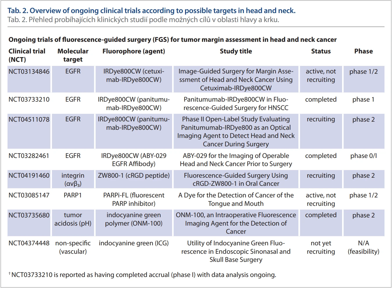
Cetuximab-IRDye800CW, a chimeric monoclonal antibody targeting the epidermal growth factor receptor (EGFR) conjugated to a near-infrared fluorophore, is among the most clinically advanced FGS agents. Given that EGFR is overexpressed in over 90% of HNSCC, cetuximab-IRDye800CW enables highly specific tumor visualization. Phase I and II clinical trials have demonstrated high tumor-to-background ratios (TBR > 5) sufficient to guide intraoperative decision-making, enabling both in situ and ex vivo assessment of surgical margins in HNSCC surgery. Reported sensitivity and specificity for detecting tumor-positive margins ranged from 85–100% [1, 53–55]. The ongoing clinical trial (NCT05499065) is evaluating the ability of cetuximab-IRDye800CW to guide the location for fresh frozen section sampling intra-operatively and to compare the detection rate of tumor-positive margins of standard histopathological analysis and fluorescence guided fresh-frozen section.
Another agent, panitumumab-IRDye 800CW, is a fully human monoclonal anti-EGFR antibody conjugated to the same near-infrared fluorophore. In early-phase clinical trials (e. g., NCT03733210 and NCT04511078), it demonstrated imaging performance comparable to cetuximab-IRDye800CW, with the added advantage of lower immunogenicity and improved safety due to its humanized structure [56, 57]. Importantly, fluorescence-based margin assessment with panitumumab-IRDye800CW achieved a sensitivity of up to 100% for detecting tumor within 2 mm of the cut surface and over 95% sensitivity and 89% specificity for identifying tumor within 5 mm [58]. This high diagnostic accuracy has been enhanced by the development of sentinel margin mapping, a technique that uses fluorescence intensity peaks on resected specimens to fastly and reliably identify the closest tumor margins [18, 59].
Beyond receptor-targeted agents, ONM-100 loaded with ICG represents a novel class of activatable fluorophores. This pH-sensitive nanoprobe fluoresces in the acidic tumor microenvironment, making it effective even in tumors with variable biomarker expression. Steinkamp et al. showed that ONM-100 identified all positive margins in a standardized surgical margin framework [60, 61].
Additional molecular targets include integrin avb3, which is upregulated in HNSCC vasculature and stroma, addressed by cRGD-ZW800-1 currently under investigation in a phase 0/I trial (NCT04191460) [56]. Similarly, PARPi-FL, a PARP1 inhibitor conjugated to a fluorophore, is being explored for tongue and oral cavity tumors (NCT03085147) [56].
Label-free methods such as dynamic optical contrast imaging (DOCI) and fluorescence lifetime imaging are also gaining traction. These approaches use endogenous fluorophore lifetimes to differentiate tissue types without the need of exogenous contrast agents [62, 63].
In summary, FGS enables real-time, high-resolution delineation of tumor margins and identification of primary lesions in head and neck surgery. Ongoing clinical trials (Tab. 2) and technological innovations continue to advance its role, including multimodal strategies that integrate tumor-specific and nerve--specific imaging, artificial intelligence--based margin analysis, and intraoperative decision support.
Ex vivo evaluation of resected specimens
In addition to in situ tumor visualization, ex vivo evaluation of resected specimens using fluorescence-guided imaging has emerged as a valuable tool for intraoperative margin assessment in HNC surgery. Traditional approaches, such as frozen section analysis, are susceptible to sampling errors, tissue deformation, and loss of orientation. In contrast, specimen-driven analysis, performed directly on the excised tumor, offers more accurate, and reproducible margin evaluation [64, 65].
Fluorescence-guided imaging of resected specimens is typically performed using closed-field systems (e. g., Pearl Trilogy, SPY Elite, or Lab-FLARE) immediately after tumor removal. Tumor-specific fluorophores like cetuximab-IRDye800CW and panitumumab-IRDye800CW have been extensively validated in this context. These agents enable precise identification of fluorescent “hot spots” of residual disease on the specimen surface, which may guide targeted frozen section sampling and immediate re-resection [18, 66].
In a pivotal study, Teraphongphom et al. demonstrated that optical mapping of head and neck surgical specimens using cetuximab-IRDye800CW enabled accurate detection of subclinical tumor foci at the resection margins, which then correlated with final histopathology results [66]. In another study by Rosenthal et al., the ex vivo fluorescence tumor mapping helped to assess tumor margins and identify the localization of positive margins [1].
A meta-analysis by Higginson et al. reported a pooled sensitivity of 95.7% and specificity of 82.7% for tumor-targeted fluorescence in margin detection, which is comparable to frozen section histology but with faster feedback and greater surface coverage [67].
While this approach requires coordination between surgical and pathology teams, it holds the potential to standardize and enhance the accuracy of margin assessment, especially in situations where visualization and surgical orientation is limited.
Sentinel and tumor-positive lymph node detection
In head and neck cancer, the presence of regional lymph node metastases is a critical prognostic factor, influencing staging, treatment planning, and overall survival. Up to 25% of patients with early-stage oral cavity or oropharyngeal squamous cell carcinoma (OSCC) harbor occult nodal metastases despite negative clinical and radiologic findings [68, 69]. While elective neck dissection remains a standard staging and therapeutic procedure, it carries a risk of functional morbidity. Sentinel lymph node biopsy has emerged as a less invasive and more selective alternative. FGS has significantly enhanced the precision of SLNB in HNC by enabling real-time intraoperative visualization of lymphatic drainage.
ICG is the most widely used near-infrared fluorophore for fluorescence-guided SLNB. When administered peritumorally, ICG migrates via lymphatic vessels to the sentinel node, which can be visualized intraoperatively. Clinical studies consistently report SLN detection rates of 92–100% using ICG alone. These findings indicate that ICG may achieve higher detection rates compared to traditional radionuclide-guided SLNB, while maintaining comparable sensitivity and specificity [70–73]. Furthermore, the use of hybrid tracers such as ICG--99mTc-nanocolloid has been shown to enhance detection rates, particularly in anatomically complex regions. In a prospective randomized trial, Stoffels et al. demonstrated significantly better SLN localization with this hybrid tracer than with radiocolloid alone [36]. Similarly, Nakamura et al. confirmed that combining ICG with radioisotopes shortened detection times and increased SLN detection [74].
Sethi et al. validated the approach of simultaneous use of gamma probes and ICG in patients with cutaneous malignancies of the head and neck, reporting reliable intraoperative mapping and high localization accuracy, including in difficult-to-access regions [34].
Beyond non-targeted agents, tumor-specific fluorophores are being evaluated for nodal mapping. Panitumumab-IRDye800CW has been shown to accurately identify both primary tumors and metastatic lymph nodes in patients with HNSCC providing high contrast imaging of metastatic nodes, with the possibility to differentiate metastatic from benign nodes based on fluorescence intensity ratios [69, 75].
In head and neck melanoma, characterized by complex and variable lymphatic drainage patterns, novel agents such as cRGDY-PEG-Cy5.5 fluorescent silica nanoparticles have been tested. In a clinical trial by Zanoni et al., these nanoparticles enabled precise SLN localization with 90% concordance to standard SLN techniques [76].
Further advances in lymph node detection include ratiometric and NIR-II multispectral techniques, which offer better tissue contrast and penetration depth. Though not yet fully translated into clinical practice, their preclinical success suggests strong future potential for visualizing nodes located deeper beneath the tissue surface [76].
Taken together, these developments suggest that fluorescence-enhanced SLNB, may improve staging accuracy and reduce unnecessary neck dissections. Ongoing clinical trials continue to assess the oncologic safety, sensitivity, and decision-making impact of fluorescence-guided lymph node mapping in head and neck surgery [2].
Applications and benefits of fluorescence-guided surgery in thyroid and parathyroid surgery
Thyroid and parathyroid surgeries are among the most frequently performed endocrine operations, but they carry significant risks, including postoperative hypocalcemia, hypoparathyroidism, and RLN injury. These complications commonly arise from inadvertent injury, devascularisation, or excision of the parathyroid glands (PGs), which are small, variably located, and difficult to distinguish from surrounding tissue. Similarly, the RLN is prone to injury due to its variable anatomy and close proximity to the thyroid gland. In this context, FGS has emerged as a valuable adjunct, offering enhanced intraoperative identification of both PGs and the RLN.
One of the most notable advancements in this field is the use of near-infrared autofluorescence (NIRAF). Parathyroid tissue naturally emits autofluorescence when excited with NIR light, enabling surgeons to identify PGs without administering exogenous contrast agents. This technique has demonstrated high accuracy, with reported sensitivities ranging from 83–100%, however, there is the high heterogeneity across clinical studies [77, 78]. A 2021 meta-analysis by Wang et al. reported overall sensitivity and specificity of 96% and 96%, respectively, for NIRAF in identifying PGs across over 2,000 patients undergoing thyroid or parathyroid surgery [78]. Its routine use has now become established in many endocrine surgery centers, particularly in total thyroidectomies and central neck dissections where the risk of PG misidentification is elevated [78, 79].
Recent randomized clinical trials have shown that NIRAF improves the PG identification rate even in low-volume centers and is beneficial in hemithyroidectomy [80]. This is especially relevant given the variability in surgical expertise across institutions.
ICG angiography complements NIRAF by providing functional assessment of PG perfusion. After intravenous administration, ICG enables real-time evaluation of PG vascularization. A meta-analysis by Kim et al. showed ICG has a sensitivity of 93.8% for PG identification and a high predictive value for postoperative function (AUC = 0.909) [79]. Di Lorenzo et al. recently demonstrated that ICG-guided surgery significantly reduced rates of both transient and permanent hypoparathyroidism after total thyroidectomy [81].
Additionally, ICG angiography has demonstrated utility in the intraoperative detection of hyperfunctioning parathyroid adenomas, which often show enhanced fluorescence due to increased vascularity. Maser et al. confirmed the feasibility of this technique in targeted parathyroidectomy [82].
Beyond PG identification, FGS shows potential for RLN visualization. Although primarily investigated in preclinical models, near-infrared ICG imaging has shown the ability to highlight richly perfused nerves such as the RLN in humans [41, 42].
The combined use of NIRAF and ICG angiography provides both structural and functional information, facilitating more accurate intraoperative decision-making. A systematic review by Barbieri et al. concluded that this multimodal approach reduces postoperative hypocalcemia more effectively than white-light visualization alone [83]. FGS is also gaining traction in minimally invasive and robotic-assisted thyroid surgeries, including transoral endoscopic thyroidectomy, where the limited surgical view can compromise PG and nerve identification [84].
Impact of fluorescence-guided surgery in clinical decision-making
FGS is transforming intraoperative decision-making in head and neck oncology by enabling real-time visualization of tumors, lymph nodes, vasculature, and nerves. This optical guidance enhances surgical precision, facilitates tailored resections, and improves both oncologic and functional outcomes.
A key benefit of FGS is its role in margin assessment. Traditional techniques such as frozen section analysis (FSA) are limited by sampling error, loss of tissue orientation, and procedural delays. In contrast, FGS provides rapid, high-resolution visualization of tumor boundaries, allowing surgeons to detect positive or close margins that may be missed with conventional methods. In a pivotal study, van Keulen et al. demonstrated that intraoperative use of panitumumab-IRDye800CW changed surgical decisions in over 21% of head and neck cancer cases, enabling detection of otherwise unrecognized tumor foci or inadequate resection margins [85]. Similarly, Steinkamp et al. showed that ONM--100, a pH-activated fluorescent agent, achieved 100% sensitivity for detecting tumor-positive margins and influenced resection extent, helping to avoid both over - and under-treatment [60].
FGS also improves lymph node mapping. Panitumumab-IRDye800CW has demonstrated high sensitivity and specificity in distinguishing metastatic from benign lymph nodes, guiding targeted pathological assessment, and influencing both the extent of neck dissection and postoperative treatment plans [69, 86, 87]. Krishnan et al. demonstrated that intravenous administration of panitumumab-IRDye800CW enabled sensitive and specific detection of both sentinel and metastatic nodes even in cases where the primary tumor was inaccessible [75]. The utility of ICG-based imaging for sentinel lymph node biopsy (SLNB) is also well documented and specificity as a non-radioactive alternative provides high detection rates and specificity [73].
The clinical utility of FGS extends to workflow efficiency. It reduces dependence on intraoperative pathology and may shorten surgical time [88]. Furthermore, a review by Srinivasan et al. confirmed that fluorescent tracers are safe, sensitive, and capable of altering intraoperative plans in a substantial number of cases [12].
Despite these benefits, some limiting factors remain. Firstly, most FGS systems currently available provide only qualitative or semi-quantitative information, relying on the surgeon’s visual interpretation of fluorescence intensity. Although efforts are ongoing to develop quantitative fluorescence imaging systems capable of displaying tumor-to-background ratios or absolute fluorophore concentration, these tools are not yet standardized or widely implemented in clinical practice.
The clinical adoption of fluorescence--guided surgery is further constrained by the limited number of fluorophores approved for human use. Targeted fluorophores, which offer higher specificity for tumor tissues and better tumour-to--background ration, remain largely investigational. Moreover, despite growing interest in the field, evidence from large, multicenter randomized controlled trials is scarce, leaving the clinical benefits of fluorescence guidance insufficiently validated.
Future perspectives in fluorescence-guided surgery
The current innovations in fluorescence--guided surgery are driven by advances in molecular imaging, artificial intelligence, and integrated optical technologies. While FGS is already transforming surgical workflows in head and neck oncology, ongoing research is steadily broadening its capabilities – moving beyond simple tumor visualization toward a more comprehensive, multi-modal surgical navigation tool with diagnostic and possibly even therapeutic applications.
One promising direction is the development of next-generation fluorophores with improved specificity, pharmacokinetics, and safety. Agents like cetuximab-IRDye800CW and panitumumab-IRDye800CW, which target EGFR in HNSCC, have demonstrated strong tumor-to-background ratios and favorable safety profiles [1, 57]. However, current research is focused on activatable probes, such as pH-sensitive and enzyme-responsive fluorophores, which fluoresce only in specific tumor environments – reducing background signal and enhancing margin delineation [60, 89].
New molecular targets are also being explored. Beyond EGFR, candidate targets include the urokinase-type plasminogen activator receptor (uPAR), folate receptor-alpha, and matrix metalloproteinases (MMPs). These molecules could expand FGS to tumors with low or variable EGFR expression [90].
Multimodal approaches, such as integrating FGS with complementary imaging technologies such as Raman spectroscopy, optical coherence tomography, and photoacoustic imaging offers a more comprehensive understanding of tissue architecture, composition, and perfusion. For example, pairing fluorescence with Raman spectroscopy can enable near-histological discrimination of tumor versus normal tissue, facilitating real-time “optical biopsy” during resection [19].
Artificial intelligence (AI) is expected to play a pivotal role in interpreting fluorescence signals. Machine learning algorithms trained on fluorescence intensity patterns can assist in intraoperative decision-making, reduce inter-surgeon variability, and provide objective thresholds for tumor margin assessment [62].
In addition to imaging, fluorescence is being explored for therapeutic applications, particularly through photodynamic therapy (PDT). Photosensitizers such as 5-aminolevulinic acid (5-ALA) and chlorin-based agents are being investigated for dual diagnostic and therapeutic use – an approach known as theranostics. These agents allow simultaneous tumor visualization and selective ablation of residual disease [91, 92].
Nevertheless, several challenges remain. One of the most significant is the limited depth penetration of near-infrared light, which typically does not exceed 1 cm in tissue. This restricts the ability to visualize deep-seated tumors or nodal metastases. Research into longer-wavelength fluorophores in the NIR-IIwindow, as well as time-gated imaging techniques, aims to overcome these physical limitations and expand the reach of fluorescence beyond superficial structures [93]. Despite these advances, no NIR-II fluorescence imaging system has yet received regulatory approval for clinical use, which currently limits its integration into routine surgical practice. Regulatory hurdles also slow the pace of clinical translation. The pathway to approval for new fluorophores is long and costly, often requiring extensive toxicological data and validation through multicenter trials. Additionally, inconsistencies in hardware performance and imaging interpretation further complicate implementation, underlining the need for industry-wide standards [68].
Conclusion
FGS has emerged as an intraoperative tool in head and neck procedures, offering real-time visualization of tumors, lymph nodes, vascular structures, and nerves. By enhancing intraoperative precision, FGS contributes to improved oncologic control and functional outcomes. The integration of both targeted fluorophores and intrinsic tissue autofluorescence has expanded its versatility in various indications. While challenges remain, such as limited depth penetration and lack of standardization, the growing body of evidence supports FGS as a tool that enhances the intraoperative decision-making in real time. As technologies continue to evolve, fluorescence imaging is expected to become an integral component of precision surgery across multiple clinical settings.
Conflict of interest statement
The author declares that there is no conflict of interest related to the topic, development, or publication of this article, and that neither its preparation nor its publication was supported by any pharmaceutical company. This declaration applies to all co-authors as well.
Funding
This work was supported by the Ministry of Health of the Czech Republic, grant No. NU21--03-00273, and by the Charles University Cooperation Program, research area Surgical Disciplines.
Zdroje
1. Rosenthal EL, Warram JM, de Boer E et al. Safety and Tumor Specificity of Cetuximab-IRDye800 for Surgical Navigation in Head and Neck Cancer. Clin Cancer Res 2015; 21 (16): 3658–3666. Doi: 10.1158/1078-0432.CCR-14-3284.
2. Crawford KL, Pacheco FV, Lee YJ et al. A Scoping Review of Ongoing Fluorescence-Guided Surgery Clinical Trials in Otolaryngology. Laryngoscope 2022; 132 (1): 36–44. Doi: 10.1002/lary.29891.
3. Drezek R, Sokolov K, Utzinger U et al. Understanding the contributions of NADH and collagen to cervical tissue fluorescence spectra: modeling, measurements, and implications. J Biomed Opt 2001; 6 (4): 385–396. Doi: 10.1117/1.1413209.
4. Wu M, Gong D, Zhou Y et al. Activatable probes with potential for intraoperative tumor-specific fluorescence-imaging guided surgery. J Mater Chem B 2023; 11 (41): 9777–9797. Doi: 10.1039/d3tb01590d.
5. Liu J, Xu Z, Meng C et al. Acidic tumor microenvironment-activatable fluorescent diagnostic probe for the rapid identification and resection of human tumors via spraying. Biosens Bioelectron 2023; 234 : 115343. Doi: 10.1016/j.bios.2023.115343
6. Zeinerová L, Černý M, Homoláč M et al. Využití fluoresceinu ke zlepšení senzitivity flexibilního endoskopického vyšetření polykání Otorinolaryngol Foniatr 2023; 72 (2): 127–135. Doi: 10.48095/ccorl2023127.
7. Archibald SJ, Holland JP, Korde A et al.. Combining Nuclear Medicine With Other Modalities: Future Prospect for Multimodality Imaging. Mol Imaging 2024; 23 : 15353508241245265. Doi: 10.1177/15353508241245265.
8. Schaafsma BE, Mieog JS, Hutteman M et al. The clinical use of indocyanine green as a near-infrared fluorescent contrast agent for image--guided oncologic surgery. J Surg Oncol 2011; 104 (3): 323–332. Doi: 10.1002/jso.21943.
9. van Leeuwen FWB, Schottelius M, Brouwer OR et al. Trending: Radioactive and Fluorescent Bimodal/Hybrid Tracers as Multiplexing Solutions for Surgical Guidance. J Nucl Med 2020; 61 (1): 13–19. Doi: 10.2967/jnumed.119.228684.
10. van Leeuwen FW, Valdes-Olmos R, Buckle T et al. Hybrid surgical guidance based on the integration of radionuclear and optical technologies. Br J Radiol 2016; 89 (1062): 20150797. Doi: 10.1259/bjr.20150797.
11. Devgan Y, Mayilvaganan S, Mishra A et al. Comparison of indocyanine green angiography vs intraoperative parathyroid hormone in early prediction of risk of post-thyroidectomy hypocalcemia: a prospective cohort study. Ann Med Surg (Lond) 2024; 86 (2): 678–688. Doi: 10.1097/MS9.0000000000001578.
12. Srinivasan A, Kaminskaite V, Winter SC. The Use of Fluorescent Markers to Detect and Delineate Head and Neck Cancer: A Scoping Review. Clin Otolaryngol 2025; 50 (2): 220–240. Doi: 10.1111/coa.14263.
13. Suero Molina E, Bruneau M, Reuter G et al. Fluorescence guidance in skull base surgery: Applications and limitations – A systematic review. Brain Spine 2024; 4 : 103328. Doi: 10.1016/j.bas.2024.103328.
14. Lee YJ, van den Berg NS, Orosco RK et al. A narrative review of fluorescence imaging in robotic-assisted surgery. Laparosc Surg 2021; 5. Doi: 10.21037/ls-20-98
15. Scott-Wittenborn N, Jackson RS. Intraoperative imaging during minimally invasive transoral robotic surgery using near-infrared light. Am J Otolaryngol 2018; 39 (2): 220–222. Doi: 10.1016/j.amjoto.2017.09.001.
16. Bergenfelz A, Barczynski M, Heie A et al. Impact of autofluorescence for detection of parathyroid glands during thyroidectomy on postoperative parathyroid hormone levels: parallel multicentre randomized clinical trial. Br J Surg 2023; 110 (12): 1824–1833. Doi: 10.1093/bjs/znad278.
17. Frey S, Bannani S, Caillard C et al. Parathyroid near-infrared autofluorescence use for parathyroidectomy in mild primary hyperparathyroidism: Results from a randomized monocentric trial. Surgery 2025; 177 : 108878. Doi: 10.1016/j.surg.2024.05.062.
18. Gao RW, Teraphongphom NT, van den Berg NS et al. Determination of Tumor Margins with Surgical Specimen Mapping Using Near-Infrared Fluorescence. Cancer Res 2018; 78 (17): 5144–5154. Doi: 10.1158/0008-5472.CAN-18-0878.
19. Lauwerends LJ, Abbasi H, Bakker Schut TC et al. The complementary value of intraoperative fluorescence imaging and Raman spectroscopy for cancer surgery: combining the incompatibles. Eur J Nucl Med Mol Imaging 2022; 49 (7): 2364–2376. Doi: 10.1007/s00259-022-05705-z.
20. Schopper S, Smeets R, Gosau M et al. Intraoperative ICG-based fluorescence-angiography in head and neck reconstruction: Predictive value for impaired perfusion of free flaps. J Craniomaxillofac Surg 2022; 50 (4): 371–379. Doi: 10.1016/j.jcms.2021.09.020.
21. Yeoh MS, Kim DD, Ghali GE. Fluorescence angiography in the assessment of flap perfusion and vitality. Oral Maxillofac Surg Clin North Am 2013; 25 (1): 61–66. Doi: 10.1016/j.coms.2012.11.004.
22. Galli A, Salerno E, Bramati C et al. Indocyanine green fluorescence video-angiography for flap perfusion assessment in head and neck reconstruction: a prospective study. Eur Arch Otorhinolaryngol 2025; 282 (2): 961–970. Doi: 10.1007/s00405-024-08959-5.
23. Beckler AD, Ezzat WH, Seth R et al. Assessment of Fibula Flap Skin Perfusion in Patients Undergoing Oromandibular Reconstruction: Comparison of Clinical Findings, Fluorescein, and Indocyanine Green Angiography. JAMA Facial Plast Surg 2015; 17 (6): 422–426. Doi: 10.1001/jamafacial.2015.0961.
24. Abdelwahab M, Kandathil CK, Most SP et al. Utility of Indocyanine Green Angiography to Identify Clinical Factors Associated With Perfusion of Paramedian Forehead Flaps During Nasal Reconstruction Surgery. JAMA Facial Plast Surg 2019; 21 (3): 206–212. Doi: 10.1001/jamafacial.2018.1829.
25. Giordano L, Galli A, Familiari M et al. Head and neck pedicled flap autonomization using a new high-resolution indocyanine green fluorescence video-angiography device. Head Neck 2022; 44 (6): 1496–1499. Doi: 10.1002/hed.27051.
26. Schlosshauer T, Rothenberger J, Heiss C et al. The role of near-infrared fluorescence imaging with indocyanine green dye in pedicle division with the paramedian forehead flap. Int Wound J 2021; 18 (6): 881–888. Doi: 10.1111/iwj.13590.
27. Surowitz JB, Most SP. Use of laser-assisted indocyanine green angiography for early division of the forehead flap pedicle. JAMA Facial Plast Surg 2015; 17 (3): 209–214. Doi: 10.1001/jamafacial.2015.0171.
28. Giordano L, Familiari M, Galli A et al. Trimming of Facial Artery Myomucosal Flap (FAMM) using Indocyanine Green Fluorescence Video-Angiography: Operative Nuances. Ann Surg Oncol 2022; 29 (13): 8361. Doi: 10.1245/s10434-022-12171-2.
29. Shaikh N, O‘Brien D, Makary C et al. Intraoperative Indocyanine Green Angiography for Assessing Flap Perfusion in Skull Base Reconstruction: A Systematic Review. J Neurol Surg B Skull Base 2022; 83 (Suppl 2): e492–e500. Doi: 10.1055/s-0041-1732309.
30. Daum R, Grimm DR, Yong M et al. Using Indocyanine Green Fluorescence Angiography to Evaluate the Impact of Surgical and Patient Factors on Posterior Septal Artery Perfusion in Endoscopic Skull Base Surgery. J Neurol Surg B Skull Base 2024. Doi: 10.1055/s-0044-1792078.
31. Noiphithak R, Yanez-Siller JC, Rukskul P et al. Indocyanine Green Angiography for Real-time Evaluation of Nasoseptal Flap Vascularity and Perfusion. Laryngoscope 2025; 135 (3): 1060–1065. Doi: 10.1002/lary.31975.
32. Singaravelu A, McCarrick C, Potter S et al. Clinical and Cost-Effectiveness of Intraoperative Flap Perfusion Assessment With Indocyanine Green Fluorescence Angiography in Breast and Head and Neck Reconstructions: A Systematic Review and Meta-Analysis. Microsurgery 2024; 44 (8): e31250. Doi: 10.1002/micr.31250.
33. Ohba S, Yokoyama J, Fujimaki M et al. Significant improvement in superselective intra-arterial chemotherapy for oral cancer by using indocyanine green fluorescence. Oral Oncol 2012; 48 (11): 1101–1105. Doi: 10.1016/j.oraloncology.2012.08.007.
34. Sethi HK, Sina EM, Mady LJ et al. Sentinel lymph node biopsy for head and neck malignancies utilizing simultaneous radioisotope gamma probe and indocyanine green fluorescence navigation. Head Neck 2024; 46 (1): 212–217. Doi: 10.1002/hed.27563.
35. Bredell MG. Sentinel lymph node mapping by indocyanin green fluorescence imaging in oropharyngeal cancer – preliminary experience. Head Neck Oncol 2010; 2 : 31. Doi: 10.1186/1758-3284-2-31.
36. Stoffels I, Leyh J, Poppel T et al. Evaluation of a radioactive and fluorescent hybrid tracer for sentinel lymph node biopsy in head and neck malignancies: prospective randomized clinical trial to compare ICG - (99m) Tc-nanocolloid hybrid tracer versus (99m) Tc-nanocolloid. Eur J Nucl Med Mol Imaging 2015; 42 (11): 1631–1638. Doi: 10.1007/s00259-015-3093-7.
37. Chao AH, Schulz SA, Povoski SP. The application of indocyanine green (ICG) and near-infrared (NIR) fluorescence imaging for assessment of the lymphatic system in reconstructive lymphaticovenular anastomosis surgery. Expert Rev Med Devices 2021; 18 (4): 367–374. Doi: 10.1080/17434440.2021.1900725.
38. Mito D, Wang YM, Wu SC et al. Use of Microscope-Integrated Near-Infrared Fluorescence for Enhancing Deep Lymphatic Vessel Detection during Supermicrosurgical Lymphaticovenous Anastomosis: A Longitudinal Cohort Study. Plast Reconstr Surg 2022; 149 (5): 1227–1233. Doi: 10.1097/PRS.0000000000009050.
39. Maus EA, Tan IC, Rasmussen JC et al. Near-infrared fluorescence imaging of lymphatics in head and neck lymphedema. Head Neck 2012; 34 (3): 448–453. Doi: 10.1002/hed.21538.
40. van Heumen S, Riksen JJM, Bramer WM et al. Imaging of the Lymphatic Vessels for Surgical Planning: A Systematic Review. Ann Surg Oncol 2023; 30 (1): 462–479. Doi: 10.1245/s10434-022-12552-7.
41. Gorobeiko M, Dinets A. Unexpected mapping of recurrent laryngeal nerve by fluorescence-guided surgery using near-infrared indocyanine green angiography. International Journal of Endocrinology (Ukraine) 2023; 19 (5): 349–353.
42. Kwon JG, Choi YJ, Kim SC et al. A technique for safe deep facial tissue dissection: Indocyanine green-assisted intraoperative real-time visualization of the vasa nervorum of facial nerve with a near-infrared camera. J Craniomaxillofac Surg 2019; 47 (11): 1819–1826. Doi: 10.1016/j.jcms.2019.07.011.
43. Haubner F, Gassner HG, Brawanski A et al. Fluorescein sodium-guided surgery of parotid gland tumors as a technical advance. BMC Ear Nose Throat Disord 2017; 17 : 6. Doi: 10.1186/s12901-017-0039-7.
44. Wang LG, Barth CW, Combs JR et al. Investigation of Oxazine and Rhodamine Derivatives as Peripheral Nerve Tissue Targeting Contrast Agent for In Vivo Fluorescence Imaging. Proc SPIE Int Soc Opt Eng 2019; 10862. Doi: 10.1117/12.2507296.
45. Wang LG, Montano AR, Masillati AM et al. Nerve Visualization using Phenoxazine-Based Near-Infrared Fluorophores to Guide Prostatectomy. Adv Mater 2024; 36 (16): e2304724. Doi: 10.1002/adma.202304724.
46. Barth CW, Shah VM, Wang LG et al. Clinically translatable formulation strategies for systemic administration of nerve-specific probes. Adv Ther (Weinh) 2021; 4 (7). Doi: 10.1002/adtp.202100002.
47. Hussain T, Mastrodimos MB, Raju SC et al. Fluorescently labeled peptide increases identification of degenerated facial nerve branches during surgery and improves functional outcome. PLoS One 2015; 10 (3): e0119600. Doi: 10.1371/journal.pone.0119600.
48. Whitney MA, Crisp JL, Nguyen LT et al. Fluorescent peptides highlight peripheral nerves during surgery in mice. Nat Biotechnol 2011; 29 (4): 352–356. Doi: 10.1038/nbt.1764.
49. Berehova N, van Meerbeek MP, Azargoshasb S et al. A Truncated 14-Amino-Acid Myelin Protein-Zero-Targeting Peptide for Fluorescence-Guided Nerve-Preserving Surgery. Biomolecules 2023; 13 (6). Doi: 10.3390/biom13060942.
50. Buckle T, Hensbergen AW, van Willigen DM et al. Intraoperative visualization of nerves using a myelin protein-zero specific fluorescent tracer. EJNMMI Res 2021; 11 (1): 50. Doi: 10.1186/s13550-021-00792-9.
51. Szafran DA, Shams NA, Montano A et al. Two--color fluorescence-guided surgery for head and neck cancer resections. J Biomed Opt 2025; 30 (Suppl 1): S13707. Doi: 10.1117/1.JBO.30.S1.S13707.
52. De Ravin E, Venkatesh S, Harmsen S et al. Indocyanine green fluorescence-guided surgery in head and neck cancer: A systematic review. Am J Otolaryngol 2022; 43 (5): 103570. Doi: 10.1016/j.amjoto.2022.103570.
53. Voskuil FJ, de Jongh SJ, Hooghiemstra WTR et al. Fluorescence-guided imaging for resection margin evaluation in head and neck cancer patients using cetuximab-800CW: A quantitative dose-escalation study. Theranostics 2020; 10 (9): 3994-4005. Doi: 10.7150/thno.43227.
54. Moore LS, Rosenthal EL, de Boer E et al. Effects of an Unlabeled Loading Dose on Tumor-Specific Uptake of a Fluorescently Labeled Antibody for Optical Surgical Navigation. Mol Imaging Biol 2017; 19 (4): 610–616. Doi: 10.1007/s11307-016-1022-1.
55. Moore LS, Rosenthal EL, Chung TK et al. Characterizing the Utility and Limitations of Repurposing an Open-Field Optical Imaging Device for Fluorescence-Guided Surgery in Head and Neck Cancer Patients. J Nucl Med 2017; 58 (2): 246–251. Doi: 10.2967/jnumed.115.171413.
56. ClinicalTrials.gov. Trials: NCT03733210, NCT04511078, NCT03282461, NCT04191460, NCT03085147.
57. Gao RW, Teraphongphom N, de Boer E et al. Safety of panitumumab-IRDye800CW and cetuximab-IRDye800CW for fluorescence-guided surgical navigation in head and neck cancers. Theranostics 2018; 8 (9): 2488–2495. Doi: 10.7150/thno.24487.
58. van Keulen S, van den Berg NS, Nishio N et al. Rapid, non-invasive fluorescence margin assessment: Optical specimen mapping in oral squamous cell carcinoma. Oral Oncol 2019; 88 : 58–65. Doi: 10.1016/j.oraloncology.2018.11.012.
59. Krishnan G, van den Berg NS, Nishio N et al. Fluorescent Molecular Imaging Can Improve Intraoperative Sentinel Margin Detection in Oral Squamous Cell Carcinoma. J Nucl Med 2022; 63 (8): 1162–1168. Doi: 10.2967/jnumed.121.262235.
60. Steinkamp PJ, Voskuil FJ, van der Vegt B et al. A Standardized Framework for Fluorescence-Guided Margin Assessment for Head and Neck Cancer Using a Tumor Acidosis Sensitive Optical Imaging Agent. Mol Imaging Biol 2021; 23 (6): 809–817. Doi: 10.1007/s11307-021-01614-z.
61. Voskuil FJ, Steinkamp PJ, Zhao T et al. Exploiting metabolic acidosis in solid cancers using a tumor-agnostic pH-activatable nanoprobe for fluorescence-guided surgery. Nat Commun 2020; 11 (1): 3257. Doi: 10.1038/s41467-020-16814-4.
62. Marsden M, Weyers BW, Bec J et al. Intraoperative Margin Assessment in Oral and Oropharyngeal Cancer Using Label-Free Fluorescence Lifetime Imaging and Machine Learning. IEEE Trans Biomed Eng 2021; 68 (3): 857–868. Doi: 10.1109/TBME.2020.3010480.
63. Weyers BW, Marsden M, Sun T et al. Fluorescence lifetime imaging for intraoperative cancer delineation in transoral robotic surgery. Transl Biophotonics 2019; 1 (1–2). Doi: 10.1002/tbio.201900017.
64. Aaboubout Y, Ten Hove I, Smits RWH et al. Specimen-driven intraoperative assessment of resection margins should be standard of care for oral cancer patients. Oral Dis 2021; 27 (1): 111–116. Doi: 10.1111/odi.13619.
65. Thomas Robbins K, Triantafyllou A, Suarez C et al. Surgical margins in head and neck cancer: Intra - and postoperative considerations. Auris Nasus Larynx 2019; 46 (1): 10–17. Doi: 10.1016/j.anl.2018.08.011.
66. Teraphongphom N, Kong CS, Warram JM et al. Specimen mapping in head and neck cancer using fluorescence imaging. Laryngoscope Investig Otolaryngol 2017; 2 (6): 447–452. Doi: 10.1002/lio2.84.
67. Higginson JA, Breik O, Thompson AH et al. Diagnostic accuracy of intraoperative margin assessment techniques in surgery for head and neck squamous cell carcinoma: A meta-analysis. Oral Oncol 2023; 142 : 106419. Doi: 10.1016/j.oraloncology.2023.106419.
68. Atallah I, Milet C, Coll JL et al. Role of near-infrared fluorescence imaging in head and neck cancer surgery: from animal models to humans. Eur Arch Otorhinolaryngol 2015; 272 (10): 2593–2600. Doi: 10.1007/s00405-014-3224-y.
69. White HW, Naveed AB, Campbell BR et al. Infrared Fluorescence-guided Surgery for Tumor and Metastatic Lymph Node Detection in Head and Neck Cancer. Radiol Imaging Cancer 2024; 6 (4): e230178. Doi: 10.1148/rycan.230178.
70. Alvarez Amezaga J, Barbier Herrero L, Pijoan del Barrio JI et al. Diagnostic efficacy of sentinel node biopsy in oral squamous cell carcinoma. Cohort study and meta-analysis. Med Oral Patol Oral Cir Bucal 2007; 12 (3): E235–E243.
71. Koual M, Benoit L, Nguyen-Xuan HT et al. Diagnostic value of indocyanine green fluorescence guided sentinel lymph node biopsy in vulvar cancer: A systematic review. Gynecol Oncol 2021; 161 (2): 436–441. Doi: 10.1016/j.ygyno.2021.01.031.
72. Thompson CF, St John MA, Lawson G et al. Diagnostic value of sentinel lymph node biopsy in head and neck cancer: a meta-analysis. Eur Arch Otorhinolaryngol 2013; 270 (7): 2115–2122. Doi: 10.1007/s00405-012-2320-0.
73. Venkatesh S, De Ravin E, Rajasekaran K. Indocyanine green fluorescence-guided sentinel lymph node biopsy: a systematic review. World J Otorhinolaryngol Head Neck Surg 2024; 15 (4): 298–307. Doi: 10.1002/wjo2.197.
74. Nakamura T, Kogashiwa Y, Nagafuji H et al. Validity of sentinel lymph node biopsy by ICG fluorescence for early head and neck cancer. Anticancer Res 2015; 35 (3): 1669–1674.
75. Krishnan G, van den Berg NS, Nishio N et al. Metastatic and sentinel lymph node mapping using intravenously delivered Panitumumab-IRDye800CW. Theranostics 2021; 11 (15): 7188–7198. Doi: 10.7150/thno.55389.
76. Li M, Zheng X, Han T et al. Near-infrared-II ratiometric fluorescence probes for non-invasive detection and precise navigation surgery of metastatic sentinel lymph nodes. Theranostics 2022; 12 (16): 7191–7202. Doi: 10.7150/thno.78085.
77. Pace-Asciak P, Russell J, Solorzano C et al. The utility of parathyroid autofluorescence as an adjunct in thyroid and parathyroid surgery 2023. Head Neck 2023; 45 (12): 3157–3167. Doi: 10.1002/hed.27538.
78. Wang B, Zhu CR, Liu H et al. The Accuracy of Near Infrared Autofluorescence in Identifying Parathyroid Gland During Thyroid and Parathyroid Surgery: A Meta-Analysis. Front Endocrinol (Lausanne) 2021; 12 : 701253. Doi: 10.3389/fendo.2021.701253.
79. Kim DH, Kim SH, Jung J et al. Indocyanine green fluorescence for parathyroid gland identification and function prediction: Systematic review and meta-analysis. Head Neck 2022; 44 (3): 783–791. Doi: 10.1002/hed.26950.
80. Abood A, Rolighed L, Ovesen T et al. Autofluorescence-guided hemithyroidectomy in a low--volume thyroid institution with no experience in parathyroid surgery: randomized clinical trial. Br J Surg 2024; 111 (4). Doi: 10.1093/bjs/znae075.
81. Di Lorenzo S, Carrillo Lizarazo JL, Dionigi G et al. Impact of near-infrared fluorescence imaging plus indocyanine green fluorescence on postoperative hypoparathyroidism rates after total thyroidectomy and central neck lymph node dissection. Br J Surg 2024; 111 (2). Doi: 10.1093/bjs/znae022.
82. Maser C, Kohlbrenner AH, Dirks R. Use of Indocyanine Green and Fluorescence Angiography in Parathyroid Surgery: A Feasibility Study. Surg Innov 2020; 27 (6): 587–593. Doi: 10.1177/1553350620956437.
83. Barbieri D, Indelicato P, Vinciguerra A et al. Autofluorescence and Indocyanine Green in Thyroid Surgery: A Systematic Review and Meta-Analysis. Laryngoscope 2021; 131 (7): 1683–1692. Doi: 10.1002/lary.29297.
84. Ouyang H, Wang B, Sun B et al. Application of Indocyanine Green Angiography in Bilateral Axillo-Breast Approach Robotic Thyroidectomy for Papillary Thyroid Cancer. Front Endocrinol (Lausanne) 2022; 13 : 916557. Doi: 10.3389/fendo.2022.916557.
85. van Keulen S, Nishio N, Fakurnejad S et al. The Clinical Application of Fluorescence-Guided Surgery in Head and Neck Cancer. J Nucl Med 2019; 60 (6): 758–763. Doi: 10.2967/jnumed.118.222810.
86. Nishio N, van den Berg NS, van Keulen S et al. Optical molecular imaging can differentiate metastatic from benign lymph nodes in head and neck cancer. Nat Commun 2019; 10 (1): 5044. Doi: 10.1038/s41467-019-13076-7.
87. Rosenthal EL, Moore LS, Tipirneni K et al. Sensitivity and Specificity of Cetuximab-IRDye800CW to Identify Regional Metastatic Disease in Head and Neck Cancer. Clin Cancer Res 2017; 23 (16): 4744–4752. Doi: 10.1158/1078-0432.CCR-16-2968.
88. Thammineedi SR, Saksena AR, Nusrath Set al. Fluorescence-guided cancer surgery-A new paradigm. J Surg Oncol 2021; 123 (8): 1679–1698. Doi: 10.1002/jso.26469.
89. Zhao J, Jin G, Weng G et al. Recent advances in activatable fluorescence imaging probes for tumor imaging. Drug Discov Today 2017; 22 (9): 1367–1374. Doi: 10.1016/j.drudis.2017.04.006.
90. Zhu L, Ma Y, Kiesewetter DO et al. Rational design of matrix metalloproteinase-13 activatable probes for enhanced specificity. ACS Chem Biol 2014; 9 (2): 510–516. Doi: 10.1021/cb400698s.
91. Kataoka H, Nishie H, Hayashi N et al. New photodynamic therapy with next-generation photosensitizers. Ann Transl Med 2017; 5 (8): 183. Doi: 10.21037/atm.2017.03.59.
92. Meulemans J, Delaere P, Vander Poorten V. Photodynamic therapy in head and neck cancer: indications, outcomes, and future prospects. Curr Opin Otolaryngol Head Neck Surg 2019; 27 (2): 136–141. Doi: 10.1097/MOO. 0000000000000521.
93. Ullah Z, Roy S, Muhammad S et al. Fluorescence imaging-guided surgery: current status and future directions. Biomater Sci 2024; 12 (15): 3765–3804. Doi: 10.1039/d4bm00410h.
Štítky
Audiologie a foniatrie Dětská otorinolaryngologie OtorinolaryngologieČlánek vyšel v časopise
Otorinolaryngologie a foniatrie
2026 Číslo 3
Nejčtenější v tomto čísle
- Diagnosis of olfactory dysfunction in chronic rhinosinusitis – a case report and literature review
- Post-COVID olfactory disorders decrease the quality of life
- Applications of fluorescence-guided surgery in head and neck surgery – a narrative review
- Minimally invasive ablation techniques for thyroid nodules