
First Czech family with paraganglioma type 1 syndrome
Authors:
A. Guha 1
 ; Martin Chovanec 1
; J. Betka 2
; T. Zelinka 3
; Z. Musil 4
; J. Bouček 2
; A. Vícha 5
; J. Plzák 2
; Martin Chovanec 1
; J. Betka 2
; T. Zelinka 3
; Z. Musil 4
; J. Bouček 2
; A. Vícha 5
; J. Plzák 2
Authors place of work:
Department of Otorhinolaryngology, Charles University, 3rd Faculty of Medicine and University Hospital Královské Vinohrady, Prague
1; Department of Otorhinolaryngology and Head and Neck surgery, Charles University, 1st Faculty of Medicine and University Hospital Motol and Homolka, Prague
2; 3rd Department of Medicine, Department of Endocrinology and Metabolism of the 1st Faculty of Medicine and General University Hospital in Prague, Prague
3; Institute of Biology and Medical Genetics of the 1st Faculty of Medicine and General University Hospital in Prague
4; Department of Pediatric Hematology and Oncology, Charles University, 2nd Faculty of Medicine and University Hospital Motol and Homolka, Prague
5
Published in the journal:
Otorinolaryngol Foniatr, 75, 2026, No. 3, pp. 238-242.
Category:
Kazuistiky
doi:
https://doi.org/10.48095/ccorl2026238
Summary
It has been well established that the familial form of head and neck paraganglioma (HNPGL) is highly associated with pathogenic variants of succinate dehydrogenase complex subunit D (SDHD) gene, that leads to Paraganglioma type 1 (PGL1) syndrome. We present the pedigree of the first family in Czech Republic diagnosed with PGL1 syndrome. A 47-year-old male patient presented to us with a carotid body paraganglioma. We detected a germline pathogenic variant p.M1V of the SDHD gene in this patient, which was also detected in two daughters. Both daughters also developed HNPGLs. Head and neck paragangliomas comprise a mere 3% of all head and neck tumors and patients remain asymptomatic for long periods. Therefore, diagnosis can easily be missed or delayed in asymptomatic cases. In conclusion, we reiterate the importance of genetic screening, tumor surveillance and tailored treatment approach in patients with germline pathogenic variants of head and neck paragangliomas.
Keywords:
HNPGL – PGL1 syndrome – SDHD pathogenic variant – genetic screening
Introduction
Head and neck paragangliomas (HNPGLs) are highly vascularised neuroendocrine tumors of paraganglial cell origin that occur in sporadic or familial forms [1]. These tumors are typically slow growing in nature and have higher prevalence among female patients [2]. Patients tend to become symptomatic between the 4th and 7th decades of life [2, 3]. Nomenclature of HNPGLs is based on the localization in the head and neck. Amongst all HNPGLs, carotid body paragangliomas (CBPGLs) represent 60% of all cases [3, 4], followed by jugulotympanic (< 35–40%) [5] and vagal (< 5%). These head and neck tumors are mostly biochemically silent, only 20% can be considered secretory [6].
Almost 40–50% of all HNPGLs are hereditary [7], comprising a significant subset without known family history. Positive family history, young age (≤ 40 years), multiple tumors and presence of carotid body tumors have a higher predilection for familial forms of the disease [2, 5, 8, 9]. The understanding of the molecular mechanism of inheritance of paragangliomas was advanced by the discovery of Succinate Dehydrogenase Subunit type D (SDHD) mutations in families with paraganglioma syndrome type 1 (PGL1) in 2000 [10]. This study also revealed the germline pathogenic variants (PVs) in the SDHD gene, located at 11q23.18. HNPGLs affect more than 90% of patients with PGL1 and multiple tumors can be found in 60 to 79% of affected patients [9, 11, 12].
Case reports
In 1999, a Czech male patient was referred to us at the Otorhinolaryngology department in General University Hospital in Prague for possible surgical management of neck tumors. The following year the younger daughter presented with discomfort in the neck. In 2002, the older daughter was also referred to us since she had similar symptoms to her father.
Clinical profile of the family
Proband
The 47-year-old male patient presented with a 1-year history of painless swelling on the left side of the neck and ringing tinnitus in the left ear. He was also recently diagnosed with hypertension. In 1993, he underwent surgical extirpation of a right-sided CBPGL and remained symptom-free for 5 years. Otherwise, he had no family history of paragangliomas/pheochromocytomas.
Otorhinolaryngological examination revealed a pulsatile resistance of 5 × 3 ×× 3 cm in the neck regions II–III, with no lymphadenopathy. On Ultrasound Duplex Carotid and CT Angiography of the neck, left-sided benign vagal PGL (VPGL) was found. Biochemical tests showed normal levels of urinary catecholamines. In 1999, he underwent extirpation of the vagal tumor.
Daughter #1
The older daughter aged 22 years complained of 3 years of painless swelling on the left side of the neck and 6 months of pulsatory tinnitus with hearing difficulty on the left side. On clinical examination an expansive mass of 4 × 3 × 3 cm could be palpated on the left side of the neck region II. Otomicroscopy showed a hypervascular blush behind the ear drum in the hypotympanum. Tympanometry was normal on the right side but recorded type B curve in the left ear. Audiometry revealed mixed hearing defect of the left ear with a predominance of the conduction component. A CBPGL was suspected on the left side on Ultrasound Duplex Carotid and CT scan of neck. MRI of the head and neck confirmed a CBPGL Shamblin III. Additionally, a T2W hypersignal was detected along the promontory. Audiological findings along with radiological investigations indicated the possible presence of a PGL of the hypotympanum. In 2003, she had surgical extirpation of the carotid body tumor along with selective neck dissection II and III. In the following year, she underwent resection of the PGL of the hypotympanum with ossiculoplasty (Total Ossicular Replacement Prosthesis). This patient did not undergo biochemical tests.
Daughter #2
The younger daughter, 19 years of age, experienced a painless growth on the right side of the neck for 5 years. On clinical examination, a resistance of 3.5 × 3 × 3 cm could be palpated on the right side of the neck region II. Otomicroscopy showed a small hypervascular expansion in the middle ear (left side) suspicious of a tympanic PGL (TPGL) with normal tympanogram and audiometric findings. Ultrasound and CT Angiography investigations confirmed the presence of a right-sided VPGL. Biochemical analysis showed normal levels of plasma catecholamines. In 2000, surgical extirpation of the vagal tumor was performed, and it was decided to monitor the left-sided TPGL due to its small size.
Genetic analysis of the family
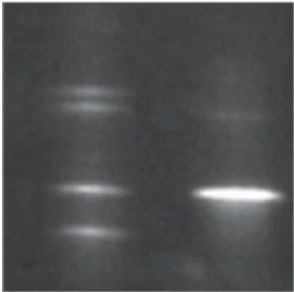
In 2004, the patient was also offered genetic testing for detection of germline pathogenic variant. He consented and peripheral blood samples were collected for this purpose. Genomic DNA was extracted from 10mL of EDTA-anticoagulated blood using standardized methods. Genetic analysis using PCR Sanger sequencing and Denaturing Gradient Gel Electrophoresis (DGGE) showed a pathogenic variant Met1Val (p.M1V) of the SDHD gene in the affected male (Fig. 1). Genetic counselling was arranged for the proband and his family explaining the necessity to test his first-degree relatives for the specified germline pathogenic variant. His two daughters were also diagnosed with the same mutation.
Outcome
On completion of genetic analysis of the family, the relevance of the genetic variant and the nature of this autosomal dominant condition was explained to the patient and his family. In 2004, after completion of surgical therapy and a series of follow-ups, the family requested to be discharged from our care as they lived in a different city. It was decided that they are to be followed up yearly at their local hospital.
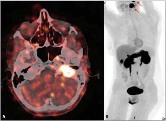
In 2024, both sisters came up for a follow-up appointment. They reported that their mother died from advanced stages of breast cancer. The older sister, 47 years of age, did not attend any regular follow--ups over the years. However, she had no new symptoms and clinically no new tumors were detected. Whilst the younger one, the 44-year-old patient complained of a 5-year history of mild tinnitus with a year of worsening of hearing on the left ear. Otomicroscopic and audiological findings of mixed hearing loss on the left side corresponded with the earlier findings of a tympanic PGL. Interestingly, CT scan of the head and neck also showed the presence of a suspected CBPGL (Fig. 2A) along with the tympanic PGL. She also underwent an 18F-DOPA PET/CT (Fig. 2B). The possibilities of surgical resection of the neck tumor and an alternative approach for the TPGL were discussed with the patient. Further information on the father was not available.
Discussion
We discuss the treatment and long-term follow-up of a family with multiple HNPGLs. Incidentally, it is also the first family that was diagnosed with PGL1 syndrome in Czech Republic. Over the last 30 years, a total of 7 HNPGLs were diagnosed in 3 family members. As mentioned before, the combination of younger patients (< 40 years of age) presenting with multiple tumors likely suggests familial disease, the latter being a hallmark for patients afflicted with SDHD PVs [7, 11, 12]. All 3 patients were under the age of 40 years when they were diagnosed with a HNPGL. They exhibited typical symptoms for the underlying findings and diagnosed tumors. Most commonly found PGLs in this family were CBPGLs and VPGLs. CBPGLs are generally of non-hereditary form in about 60% of the patients [5]. However, the presence of carotid body tumor, whether in solitary or bilateral form should be considered a risk for germline SDHD disease [9, 11].
In our family, the proband had a negative family history of paragangliomas/ pheochromocytomas. According to many studies, the percentage of germline SDHD PVs with positive family history could be as high as 80% to 100% [12–16]. In contrast, our recently published article studying Czech patients showed only one-third of Czech patients with known family history showed germline SDHD PVs [17]. There is a fair percentage of patients that are considered “occult familial cases”, the term being used when a germline mutation is discovered in an apparently sporadic case with negative family history [18], our proband represents such a case. This is not an uncommon finding in Czech Republic [17].
The inheritance pattern in this PGL1 is also noteworthy, PGL1 families exhibit an atypical inheritance pattern with a distinct “parent-of-origin dependent effect” [19]. Although SDHD PVs can be inherited both via the paternal and maternal lineage, PGLs develop after maternal transmission only in extremely rare cases [8, 20]. In 50% of the cases, maternally derived SDHD mutation carriers pass the mutation to their offspring, hence PGL1 can seem to skip generations, which may in part explain the occurrence of the disease in apparently nonfamilial cases. However, an individual who inherits an SDHD PV from his/her father is at a high risk of developing PGLs [21], an observation which was clearly made in our 2 patients. Genetic sequencing analysis found SDHD M1V (c.1A>G) germline PV in all 3 patients, thus affecting the initiation codon, that is usually disease-related when both alleles are affected [22]. Although HNPGLs are commonly seen with p.M1V PV, it has been related to early-onset functional bilateral pheochromocytomas, hence patients are at risk.
The main objectives in patients with multiple HNPGLs and/or familial disease are tailored management with effective tumor surveillance, facilitating timely treatment and achieving disease control. Tumor surveillance includes whole body imaging, biochemical analysis and effective genetic counselling and testing [23]. In HNPGLs, surgery is the mainstay therapy. With the advent of preoperative embolization, rate of perioperative neurovascular morbidity and mortality has been reduced drastically. Although there is a risk of cranial nerve injury and can be debilitating in patients, it is dependent on the localization and size of the tumor as well as surgeon’s skills. Curative rates of complete surgical removal of carotid, vagal and tympanic PGLs are between 90 and 100% [4, 24, 25], and about 78% in jugular PGLs [24]. Therefore, in multiple HNPGLs, it is recommended not to perform single stage surgery, instead the largest tumor should be removed first and in contralateral tumors, other therapeutic approaches in combination with surgery should be considered [23]. Radiotherapy is the 2nd most common therapeutic approach since PGLs are radiosensitive, however there is a lack of consensus and evidence on efficacy on growing HNPGLs. This technique reduces the risk of cranial nerve morbidities associated with surgery. Radiosurgery is preferred over conventional radiotherapy as it provides more precise therapy and has fewer side effects than the latter approach. The 10-year progression-free survival using radiosurgery can be above 90% [26]. Radionuclide therapy, therapeutic radiation and targeted therapy are based largely on the mechanism of tumorigenesis in HNPGLs [7], and while they hold a promising future, they can currently be used only in a limited number of cases with more specific criteria. Lastly, it should be mentioned wait and scan approach has been popularized since PGLs are often slow growing. It is a conservative approach that can be used on a long-term basis to monitor asymptomatic cases, small stable tumors, and patients with low risk of malignancy. This approach reduces the risk of comorbidities related to surgery or radiosurgery, but extreme caution should be exercised in multiple HNPGLs with germline PVs, there is a risk of damaging deeper structures leading to irreparable complications [27].
Conclusion
We presented a family with PGL1 syndrome and multiple HNPGLs. Patients should understand the course of the disease, as they have a lifelong risk of developing new tumors. We recommend a tailor-made approach for patients with germline PVs and multiple tumors. Management should include genetic screening for suspected carriers, tumor surveillance through meticulous follow-ups and therapeutic approaches that maximizes disease control whilst minimizing debilitating effects on the patients.
Conflict of interest
The authors declare that the research was conducted in the absence of any commercial or financial relationships that could be construed as a potential conflict of interest.
Grant support
This work was supported by the Cooperatio Program, research area SURG.
Acknowledgements
Institute of Nuclear Medicine, 1st Faculty of Medicine, Charles University and General University Hospital in Prague.
We would also like to kindly thank all our clinical and laboratory staff who supported us in our research. We are also very grateful to the patients who have participated in this study.
Zdroje
1. Mete O, Wenig BM. Update from the 5th Edition of the World Health Organization Classification of Head and Neck Tumors: overview of the 2022 WHO classification of head and neck neuroendocrine neoplasms. Head Neck Pathol 2022; 16 (1): 123–142. Doi: 10.1007/s12 105-022-01435-8.
2. Guha A, Musil Z, Vicha A et al. A systematic review on the genetic analysis of paragangliomas: primarily focused on head and neck paragangliomas. Neoplasma 2019; 66 (5): 671–680. Doi: 10.4149/neo_2018_181208N933.
3. Boedeker CC. Paragangliomas and paraganglioma syndromes. GMS Curr Top Otorhinolaryngol Head Neck Surg 2011; 10: Doc03. Doi: 10.3205/cto000076.
4. Robertson V, Poli F, Hobson B et al. A systematic review and meta-analysis of the presentation and surgical management of patients with carotid body tumours. Eur J Vasc Endovasc Surg 2019; 57 (4): 477–486. Doi: 10.1016/j.ejvs.2018.10.038.
5. Smith JD, Harvey RN, Darr OA et al. Head and neck paragangliomas: a two-decade institutional experience and algorithm for management. Laryngoscope Investig Otolaryngol 2017; 2 (6): 380–389. Doi: 10.1002/lio2.122.
6. van Duinen N, Corssmit EPM, de Jong WHA et al. Plasma levels of free metanephrines and 3-methoxytyramine indicate a higher number of biochemically active HNPGL than 24-h urinary excretion rates of catecholamines and metabolites. Eur J Endocrinol 2013; 169 (3): 377–382. Doi: 10.1530/EJE-13-0529.
7. Taïeb D, Wanna GB, Ahmad M et al. Clinical consensus guideline on the management of phaeochromocytoma and paraganglioma in patients harbouring germline SDHD pathogenic variants. Lancet Diabetes Endocrinol 2023; 11 (5): 345–361. Doi: 10.1016/S2213-8587 (23) 00038-4.
8. Boedeker CC, Hensen EF, Neumann HPH et al. Genetics of hereditary head and neck paragangliomas. Head Neck 2014; 36 (6): 907–916. Doi: 10.1002/hed.23436.
9. Neumann HPH, Erlic Z, Boedeker CC et al. Clinical predictors for germline mutations in head and neck paraganglioma patients: cost reduction strategy in genetic diagnostic process as fall-out. Cancer Res 2009; 69 (8): 3650–3656. Doi: 10.1158/0008-5472.CAN-08-4057.
10. Baysal BE, Ferrell RE, Willett-Brozick JE et al. Mutations in SDHD, a mitochondrial complex II gene, in hereditary paraganglioma. Science 2000; 287 (5454): 848–851. Doi: 10.1126/science.287.5454.848.
11. Burnichon N, Rohmer V, Amar L et al. The succinate dehydrogenase genetic testing in a large prospective series of patients with paragangliomas. J Clin Endocrinol Metab 2009; 94 (8): 2817–2827. Doi: 10.1210/jc.2008-2504.
12. Dannenberg H, Dinjens WNM, Abbou M et al. Frequent germ-line succinate dehydrogenase subunit D gene mutations in patients with apparently sporadic parasympathetic paraganglioma. Clin Cancer Res 2002; 8 (7): 2061–2066.
13. Astuti D, Hart-Holden N, Latif F et al. Genetic analysis of mitochondrial complex II subunits SDHD, SDHB and SDHC in paraganglioma and phaeochromocytoma susceptibility. Clin Endocrinol (Oxf) 2003; 59 (6): 728–733. Doi: 10.1046/j.1365-2265.2003.01914.x.
14. Fakhry N, Niccoli-Sire P, Barlier-Seti A et al. Cervical paragangliomas: is SDH geneticanalysis systematically required? Eur Arch Otorhinolaryngol 2008; 265 (5): 557–563. Doi: 10.1007/s00405-007-0517-4.
15. Papaspyrou K, Mewes T, Rossmann H et al. Head and neck paragangliomas: report of 175 patients (1989–2010). Head Neck 2012; 34 (5): 632–637. Doi: 10.1002/hed.21790.
16. Piccini V, Rapizzi E, Bacca A et al. Head and neck paragangliomas: genetic spectrum and clinical variability in 79 consecutive patients. Endocr Relat Cancer 2012; 19 (2): 149–155. Doi: 10.1530/ERC-11-0369.
17. Guha A, Vicha A, Zelinka T et al. High incidence of occult familial SDHD cases amongst Czech patients with head and neck paragangliomas. Front Endocrinol (Lausanne) 2023; 14 : 1278175. Doi: 10.3389/fendo.2023.1278175.
18. Heesterman BL, Bayley JP, Tops CM et al. High prevalence of occult paragangliomas in asymptomatic carriers of SDHD and SDHB gene mutations. Eur J Hum Genet 2013; 21 (4): 469–470. Doi: 10.1038/ejhg.2012.203.
19. van Schothorst EM, Jansen JC, Grooters E et al. Founder effect at PGL1 in hereditary head and neck paraganglioma families from the Netherlands. Am J Hum Genet 1998; 63 (2): 468–473. Doi: 10.1086/301951.
20. Pigny P, Vincent A, Cardot Bauters C et al. Paraganglioma after maternal transmission of a succinate dehydrogenase gene mutation. J Clin Endocrinol Metab 2008; 93 (5): 1609–1615. Doi: 10.1210/jc.2007-1989.
21. Else T, Greenberg S, Fishbein L. Hereditary paraganglioma-pheochromocytoma syndromes. In: Adam MP, Bick S, Mirzaa GM et al (eds). GeneReviews® [Internet]. Seattle (WA): University of Washington, Seattle; 1993–2026.
22. Riemann K, Sotlar K, Kupka S et al. Chromosome 11 monosomy in conjunction with a mutated SDHD initiation codon in nonfamilial paraganglioma cases. Cancer Genet Cytogenet 2004; 150 (2): 128–135. Doi: 10.1016/j.cancergencyto.2003.10.013.
23. Guha A, Vicha A, Zelinka T et al. Genetic variants in patients with multiple head and neck paragangliomas: dilemma in management. Biomedicines 2021; 9 (6): 626. Doi: 10.3390/biomedicines9060626.
24. Suárez C, Rodrigo JP, Bödeker CC et al. Jugular and vagal paragangliomas: systematic study of management with surgery and radiotherapy. Head Neck 2013; 35 (8): 1195–1204. Doi: 10.1002/hed.22976.
25. Forest JA jr, Jackson CG, McGrew BM. Long-term control of surgically treated glomus tympanicum tumors. Otol Neurotol 2001; 22 (2): 232–236. Doi: 10.1097/00129492-200103000-00020.
26. Krych AJ, Foote RL, Brown PD et al. Long--term results of irradiation for paraganglioma. Int J Radiat Oncol Biol Phys 2006; 65 (4): 1063–1066. Doi: 10.1016/j.ijrobp.2006.02.020.
27. Jansen TTG, Timmers HJLM, Marres HAM et al. Feasibility of a wait-and-scan period as initial management strategy for head and neck paraganglioma. Head Neck 2017; 39 (10): 2088–2094. Doi: 10.1002/hed.24871.
Štítky
Audiologie a foniatrie Dětská otorinolaryngologie OtorinolaryngologieČlánek vyšel v časopise
Otorinolaryngologie a foniatrie
2026 Číslo 3
Nejčtenější v tomto čísle
- Diagnosis of olfactory dysfunction in chronic rhinosinusitis – a case report and literature review
- Post-COVID olfactory disorders decrease the quality of life
- Applications of fluorescence-guided surgery in head and neck surgery – a narrative review
- Minimally invasive ablation techniques for thyroid nodules